Main ContentConfocal Microscopy
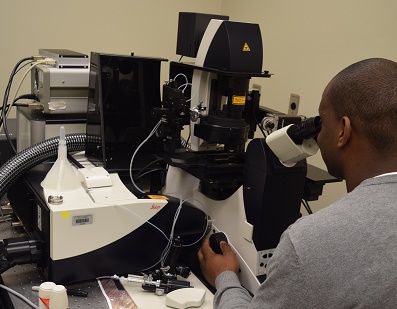
The Confocal Core Facility is located on the second floor of the new Arthur C. Guyton Research Building in Room G272. The Confocal Core houses a Leica TCS-SP2 laser scanning confocal upright microscope and a Leica TCS-SP8 multiphoton confocal microscope. Both confocal systems have three separate lasers for use with 3 standard visible length fluorescence dyes as well as transmitted light. The multiphoton includes a separate infrared laser.
Confocal Core Services
The SP2 confocal microscope equipped with three individual lasers (488/546/633 nm) capable of imaging standard fluorescence dyes within the visible spectra, including FITC/TRITC, CY2/CY3/CY5, green fluorescence protein variants (GFP/YFP/RFP/DsRed) and many different indicator dyes (i.e. Ca2+, pH, membrane potential, oxidative stress, etc). This system includes an upright microscope.
The SP8 is equipped with three individual lasers (488/561/633 nm) capable of imaging standard fluorescence dyes within the visible spectra. This system uses a light green laser (561) instead of the 546 green laser. The 561 laser can still be used with standard CY3 and TRITC dyes, but Alexa 555 dyes are ideal. The multiphoton capability is provided by a Coherent sapphire laser. This system includes an inverted microscope with a Luden environmental chamber for temperature control.
 Capabilities
- Epifluorescence provided by a mercury lamp (SP2) or halogen lamp (SP8)
- Both SP2 and SP8 include motorized stages
- Standard confocal microscopy with simultaneous or sequential image collection
- Line scan, Z-scan modes
- High speed resonant scanner
- High resolution imaging
- 10X, 20X, 40X, 40X oil, 60X oil and 100X oil objectives
- Transmitted light detection
- Tunable collection filter
- Time lapse
- Stage adaptable for slides, dishes up to 35mm, 25mm glass cover slip in an atto chamber
- Image enhancement
- Quantitation, 3D reconstruct
- High speed resonant scanner
- High resolution image
- Objectives: 63X glycerol, 1.37 NA, 40X oil 1.25 NA, 20X dry 0.7 NA, 10X dry
- Super Z galvo stage
- 6PMTS: 3 color imaging (includes 2 high sensitivity hybrid detector, standard PMT), 1 transmitted light PMT, 2 non-descanned light detectors
- Inverted orientation permits fixed or live cell/tissue imaging.
- FRET/FRAP (Fluorescence resonance excitation transfer/ Fluorescence recovery after photobleaching)
- Quantitation and colocalization
- 3D Reconstruction
- Multiphoton imaging (deep tissue imaging, dark/dense tissue far red imaging)
Fees
- Training: $100/hr
- User-based imaging: $50/hr
The Confocal Microscopy Core is primarily an equipment core. Users new to confocal microscopy typically require 2-4 hours of training depending on level of microscopy experience. Experienced users typically require 1-2 hours of training.
- All users must be trained and pre-approved prior to use by the Confocal Core director.
For more information